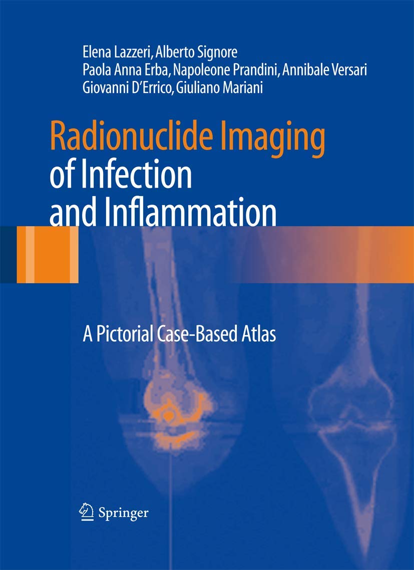
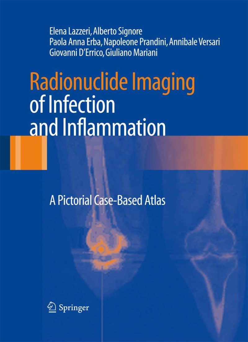
تصویربرداری رادیونوکلئیدی از عفونت و التهاب: اطلس تصویری مبتنی بر مورد ۲۰۱۲
Radionuclide Imaging of Infection and Inflammation: A Pictorial Case-Based Atlas 2012
دانلود کتاب تصویربرداری رادیونوکلئیدی از عفونت و التهاب: اطلس تصویری مبتنی بر مورد ۲۰۱۲ (Radionuclide Imaging of Infection and Inflammation: A Pictorial Case-Based Atlas 2012) با لینک مستقیم و فرمت pdf (پی دی اف)
| نویسنده |
Alberto Signore, Annibale Versari, Elena Lazzeri, Giovanni D ́Errico, Giuliano Mariani, Napoleone Prandini, Paola Anna Erba |
|---|
ناشر:
Springer Milan

۳۰ هزار تومان تخفیف با کد «OFF30» برای اولین خرید
| سال انتشار |
2012 |
|---|---|
| زبان |
English |
| تعداد صفحهها |
331 |
| نوع فایل |
|
| حجم |
26 Mb |
🏷️ 200,000 تومان قیمت اصلی: 200,000 تومان بود.129,000 تومانقیمت فعلی: 129,000 تومان.
🏷️
378,000 تومان
قیمت اصلی: ۳۷۸٬۰۰۰ تومان بود.
298,000 تومان
قیمت فعلی: ۲۹۸٬۰۰۰ تومان.
📥 دانلود نسخهی اصلی کتاب به زبان انگلیسی(PDF)
🧠 به همراه ترجمهی فارسی با هوش مصنوعی
🔗 مشاهده جزئیات
دانلود مستقیم PDF
ارسال فایل به ایمیل
پشتیبانی ۲۴ ساعته
توضیحات
معرفی کتاب تصویربرداری رادیونوکلئیدی از عفونت و التهاب: اطلس تصویری مبتنی بر مورد ۲۰۱۲
این اطلس با مستندسازی دقیق نقش تصویربرداری پزشکی هسته ای از عفونت و التهاب، خلا موجود در منابع علمی را پر می کند. سازوکارهای پاتوفیزیولوژیک و مولکولی که مبنای تصویربرداری رادیونوکلئیدی از عفونت/التهاب هستند به وضوح شرح داده شده اند، اما تمرکز اصلی کتاب بر اهمیت بالینی این روش ها است. تاثیر آن ها با مجموعه ای از موارد آموزشی غنی و مصور به نمایش گذاشته می شود که الگوهای اسکنتیگرافی رایج، و همچنین واریانت های آناتومیکی و خطاهای فنی را توصیف می کنند. توجه کافی به کاربرد تکنیک های جدیدتر، از جمله تصویربرداری فیوژن چندوجهی مانند SPECT/CT و PET/CT معطوف شده است. به ویژه، بر توانایی تصویربرداری چندوجهی در افزایش هم حساسیت و هم ویژگی تصویربرداری رادیونوکلئیدی تاکید شده است. این اطلس ابزار یادگیری بسیار خوبی برای رزیدنت های پزشکی هسته ای خواهد بود و برای سایر متخصصان علاقه مند به این حوزه روشنگر خواهد بود.
توضیحات(انگلیسی)
This atlas fills a gap in the literature by documenting in detail the role of nuclear medicine imaging of infection and inflammation. The pathophysiologic and molecular mechanisms on which radionuclide imaging of infection/inflammation is based are clearly explained, but the prime focus of the book is on the clinical relevance of such procedures. Their impact is demonstrated by a collection of richly illustrated teaching cases that describe the most commonly observed scintigraphic patterns, as well as anatomic variants and technical pitfalls. Due attention is paid to the application of recently developed techniques, including multimodality fusion imaging such as SPECT/CT and PET/CT. Emphasis is placed in particular on the ability of multimodality imaging to increase both the sensitivity and the specificity of radionuclide imaging. This atlas will be an excellent learning tool for residents in nuclear medicine and illuminating for other specialists with an interest in the field.
دیگران دریافت کردهاند
درمان سرطان موضعی-ناحیه ای با رادیونوکلئید: جنبه های بالینی و علمی ۲۰۲۰
Locoregional Radionuclide Cancer Therapy: Clinical and Scientific Aspects 2020
بیوشیمی پزشکی, پزشکی, پزشکی بالینی, پیراپزشکی, رادیولوژی، رادیوتراپی و پزشکی هسته ای, طب داخلی, فناوری های تصویربرداری
🏷️ 200,000 تومان قیمت اصلی: 200,000 تومان بود.129,000 تومانقیمت فعلی: 129,000 تومان.
تصویربرداری پاراتیروئید با رادیونوکلئید: کتاب و اطلس ۲۰۱۹
Radionuclide Parathyroid Imaging: Book and Atlas 2019
🏷️ 200,000 تومان قیمت اصلی: 200,000 تومان بود.129,000 تومانقیمت فعلی: 129,000 تومان.
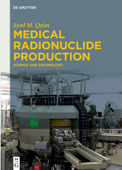
تولید رادیونوکلئیدهای پزشکی: علم و تکنولوژی ۲۰۱۹
Medical Radionuclide Production: Science and Technology 2019
🏷️ 200,000 تومان قیمت اصلی: 200,000 تومان بود.129,000 تومانقیمت فعلی: 129,000 تومان.
رادیونوکلئیدها در محیط زیست ۲۰۱۳
Radionuclides in the Environment 2013
🏷️ 200,000 تومان قیمت اصلی: 200,000 تومان بود.129,000 تومانقیمت فعلی: 129,000 تومان.
تصویربرداری استخوان با رادیونوکلئید و روش های ترکیبی ۲۰۱۳
Radionuclide and Hybrid Bone Imaging 2013
🏷️ 200,000 تومان قیمت اصلی: 200,000 تومان بود.129,000 تومانقیمت فعلی: 129,000 تومان.
رادیونوکلئیدها در محیط زیست: کنفرانس بینالمللی ایزوتوپها در مطالعات محیطی: انجمن آبزیان ۲۰۰۴، ۲۵-۲۹ اکتبر، موناکو ۲۰۰۶
Radionuclides in the Environment: International Conference on Isotopes in Environmental Studies : Aquatic Forum 2004, 25-29 October, Monaco 2006
🏷️ 200,000 تومان قیمت اصلی: 200,000 تومان بود.129,000 تومانقیمت فعلی: 129,000 تومان.
✨ ضمانت تجربه خوب مطالعه
بازگشت کامل وجه
در صورت مشکل، مبلغ پرداختی بازگردانده می شود.
دانلود پرسرعت
دانلود فایل کتاب با سرعت بالا
ارسال فایل به ایمیل
دانلود مستقیم به همراه ارسال فایل به ایمیل.
پشتیبانی ۲۴ ساعته
با چت آنلاین و پیامرسان ها پاسخگو هستیم.
ضمانت کیفیت کتاب
کتاب ها را از منابع معتیر انتخاب می کنیم.

