-100%

دانلود کتاب اطلس آناتومی گری ۲۰۲۰ (Gray’s Atlas of Anatomy 2020) با لینک مستقیم و فرمت pdf (پی دی اف)
| نویسنده |
A. Wayne Vogl, Adam W. M. Mitchell, Paul Richardson, Richard Drake, Richard Tibbitts |
|---|

۳۰ هزار تومان تخفیف با کد «OFF30» برای اولین خرید
| سال انتشار |
2020 |
|---|---|
| زبان |
English |
| تعداد صفحهها |
648 |
| نوع فایل |
|
| حجم |
144 Mb |
200,000 تومان قیمت اصلی: 200,000 تومان بود.0 تومانقیمت فعلی: 0 تومان.
🏷️
378,000 تومان
قیمت اصلی: ۳۷۸٬۰۰۰ تومان بود.
298,000 تومان
قیمت فعلی: ۲۹۸٬۰۰۰ تومان.
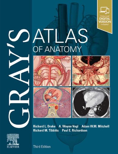
اطلس آناتومی گری، یک منبع بالینی، با تصاویر واضح و منسجم، و سازماندهی منطقی، و همراهی برای کتاب محبوب *آناتومی گری برای دانشجویان*، تصویری روشن و دیدنی از ساختارهای آناتومیکی ارائه می دهد. تصاویر خیره کننده، ارتباط ساختارها با تصاویر بالینی و آناتومی سطحی را نشان می دهند – که برای شناسایی صحیح در آزمایشگاه تشریح و آمادگی موفقیت آمیز برای امتحانات درس ضروری است.
* دانش آناتومی موجود خود را با ساختارهایی که از سطحی به عمقی ارائه می شوند، بنا کنید و نشان دهنده یک پیشرفت منطقی در سراسر بدن باشید.
* ساختارهای مختلف آناتومیکی بدن را شناسایی کنید و روابط آنها را با یکدیگر با راهنمایی بصری نزدیک به ۱۰۰۰ شکل آناتومیکی نفیس بهتر درک کنید.
* ارتباط بالینی بین ساختارهای آناتومیکی و نشانه های سطحی را با عکس های آناتومی سطحی که با نقاشی های آناتومیکی همپوشانی دارند تجسم کنید.
* ساختارهای آناتومیکی را همانطور که در عمل ارائه می شوند از طریق بیش از ۲۷۰ تصویر بالینی – از جمله تصاویر لاپاروسکوپی، رادیولوژیکی، جراحی، افتالموسکوپی، اتوسکوپی، و سایر نماهای بالینی – که در کنار آثار هنری آناتومیکی برای مقایسه دو به دو قرار داده شده اند، تشخیص دهید.
* از طریق یک تصویر جدید با فرمت بزرگ، و همچنین تصاویر جدید که آناتومی را همانطور که در محیط بالینی مدرن مشاهده می شود، منعکس می کنند، درک کامل تری از ناحیه اینگوینال در زنان به دست آورید.
* سایت آموزشگر Evolve با یک مجموعه تصویر و ویدیو در اختیار اساتید از طریق نماینده فروش Elsevier یا از طریق درخواست در https://evolve.elsevier.com قرار دارد.
Clinically focused, consistently and clearly illustrated, and logically organized, Gray’s Atlas of Anatomy, the companion resource to the popular Gray’s Anatomy for Students, presents a vivid, visual depiction of anatomical structures. Stunning illustrations demonstrate the correlation of structures with clinical images and surface anatomy – essential for proper identification in the dissection lab and successful preparation for course exams.
✨ ضمانت تجربه خوب مطالعه
در صورت مشکل، مبلغ پرداختی بازگردانده می شود.
دانلود فایل کتاب با سرعت بالا
دانلود مستقیم به همراه ارسال فایل به ایمیل.
با چت آنلاین و پیامرسان ها پاسخگو هستیم.
کتاب ها را از منابع معتیر انتخاب می کنیم.