Sale

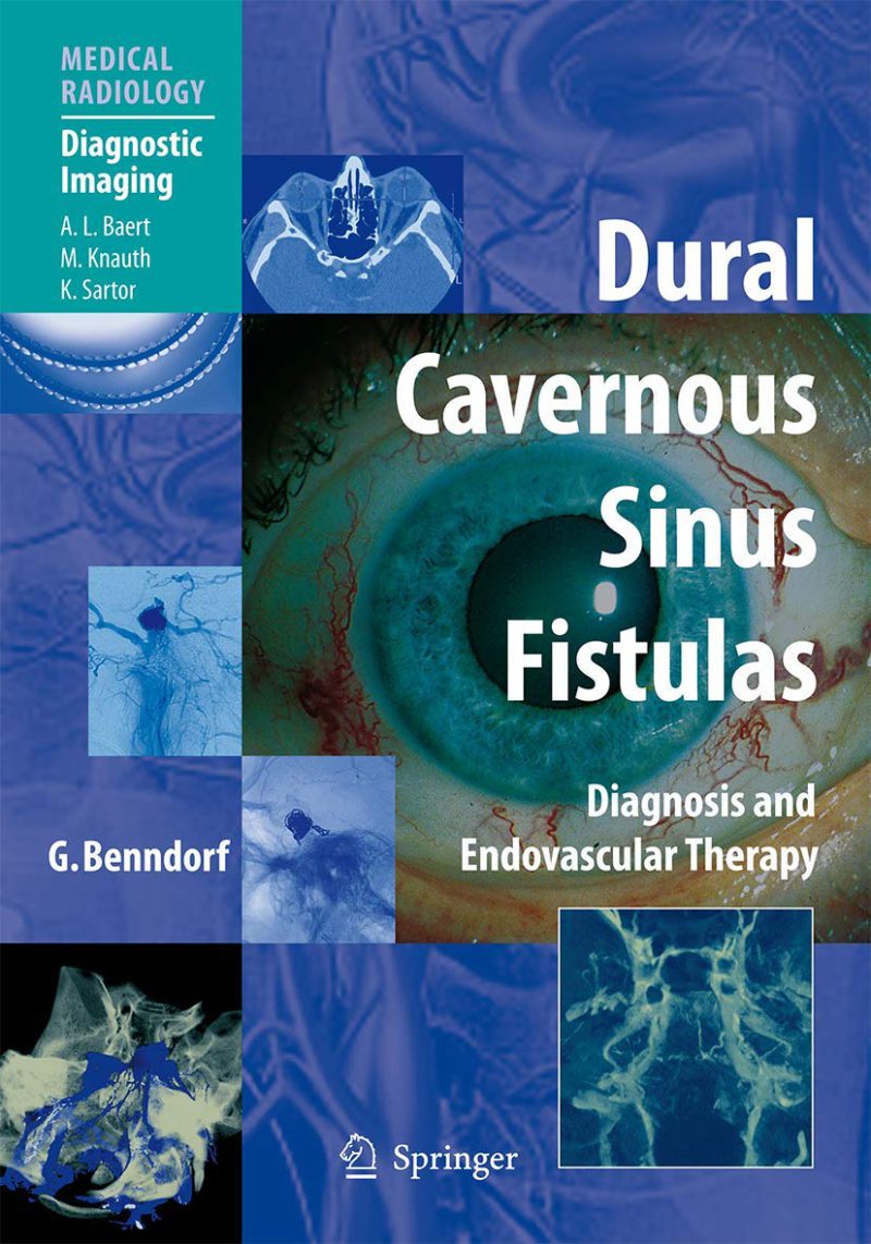
دانلود کتاب فیستول های دورال سینوس کاورنوس: تشخیص و درمان اندوواسکولار ۲۰۰۹ (Dural Cavernous Sinus Fistulas: Diagnosis and Endovascular Therapy 2009) با لینک مستقیم و فرمت pdf (پی دی اف)
| نویسنده |
Goetz Benndorf |
|---|

۳۰ هزار تومان تخفیف با کد «OFF30» برای اولین خرید
| سال انتشار |
2009 |
|---|---|
| زبان |
English |
| تعداد صفحهها |
320 |
| نوع فایل |
|
| حجم |
22 Mb |
🏷️ 200,000 تومان قیمت اصلی: 200,000 تومان بود.129,000 تومانقیمت فعلی: 129,000 تومان.
🏷️
378,000 تومان
قیمت اصلی: ۳۷۸٬۰۰۰ تومان بود.
298,000 تومان
قیمت فعلی: ۲۹۸٬۰۰۰ تومان.
Dural cavernous sinus fistulas (DCSFs) are benign vascular diseases consisting in an arteriovenous shunt at the cavernous sinus that if misdiagnosed can lead to potentially serious ophthalmologic complications. This volume provides a complete guide to the diagnosis and minimal invasive treatment of DCSFs. After sections on anatomy and classification, etiology and pathogenesis of DCSFs, the symptomatology of the disease is described in detail. The role of modern imaging techniques in the diagnosis of DCSFs is then addressed. Digital subtraction angiography (DSA) remains the gold standard for clinical decision-making; here, full consideration is given to both, conventional 2D DSA and rotational 3D angiography. Recent technological advances in this field such as Dual Volume (DV) imaging and angiographic computed tomography (ACT) are considered as well. Due attention is further paid to the use of computed tomography, magnetic resonance imaging and ultrasound. Finally, the therapeutic management of DCSFs with emphasis on various transvenous occlusion techniques are discussed in depth. This well-illustrated volume will be invaluable to all who may encounter DCSF in their clinical practice.
✨ ضمانت تجربه خوب مطالعه
در صورت مشکل، مبلغ پرداختی بازگردانده می شود.
دانلود فایل کتاب با سرعت بالا
دانلود مستقیم به همراه ارسال فایل به ایمیل.
با چت آنلاین و پیامرسان ها پاسخگو هستیم.
کتاب ها را از منابع معتیر انتخاب می کنیم.