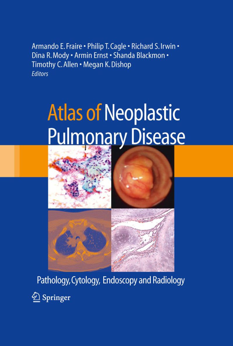
معرفی کتاب اطلس بیماری های نئوپلاستیک ریه: آسیب شناسی، سیتولوژی، اندوسکوپی و رادیولوژی ۲۰۱۰
تشخیص سرطان ریه و تومورهای خوش خیم ریوی می تواند چالش برانگیز باشد. این تشخیص می تواند از طریق مطالعه ی تصاویری تسهیل شود که امکان تشخیص الگوهای بیماری را، هم در سطوح بالینی و هم در سطوح آسیب شناسی، فراهم می کنند. از نظر مفهومی، اطلس ها کتاب های تخصصی هستند که به شدت بر تصاویر تکیه دارند تا موضوعات مختلف را به تصویر بکشند. مطابق با چنین مفهومی، این اطلس برای پر کردن خلا در رویکرد تشخیص توسعه یافته است. بر خلاف اطلس های مرسوم قبلی، این اطلس از این جهت منحصر به فرد است که تصاویر از چهار رشته اصلی (آندوسکوپی، رادیولوژی، بافت شناسی و سیتوپاتولوژی) که در مطالعه و تشخیص تومورهای ریه دخیل هستند، در یک جلد واحد گردآوری شده اند. اطلس به 11 بخش شامل 41 فصل سازماندهی شده است و از طبقه بندی 2004 تومورهای ریه توسط سازمان بهداشت جهانی (WHO) تبعیت می کند. بر این اساس، فصل ها طیف گسترده ای از موجودیت های نئوپلاستیک ریه را نشان می دهند. این اطلس با تومورهای کودکان شروع می شود و پس از آن بخش هایی در مورد تومورهای خوش خیم اپیتلیال، تومورهای غدد بزاقی، نئوپلاسم های مزانشیمی، اختلالات لنفوپرولیفراتیو، تومورهای کارسینوئید و بخشی از تومورهای متفرقه وجود دارد. تصاویری که به نظر ما بهترین نماینده ی تومورها هستند در اینجا ارائه شده اند. در مواردی، ما همکاری و مواد را از سایر متخصصان در این زمینه جمع آوری کرده ایم.
توضیحات(انگلیسی)
The diagnosis of lung cancer and benign pulmonary put forward images that in our opinion best represent tumors can be challenging. This diagnosis can be facil- the tumor entities. In some instances, we have recruited itated by the study of images that allow recognition of the collaboration and materials from other workers in the patterns of disease, both at the clinical and pathologic ?eld. levels. Conceptually de?ned, atlases are specialized books The atlas is organized into 11 parts containing 41 that rely heavily upon images to illustrate any subject mat- chapters, closely following the 2004 Classi?cation of ter. Fitting with such a concept, this atlas was developed Lung Tumors by the World Health Organization (WHO). to ?ll a void in the approach to diagnosis. In contrast to Accordingly, the chapters represent a wide range of n- previous conventional atlases, this atlas is unique in that plastic lung entities. It begins with tumors of children images from four major disciplines (endoscopy, radiology, followed by sections on benign epithelial tumors, salivary histopathology, and cytopathology) involved in the study gland tumors, mesenchymal neoplasms, lymphoprol- and diagnosis of lung tumors are brought together in a erative disorders, cardcinoid tumors, and a section of single volume. miscellaneous tumors.