مشاهده سبد خرید “Minimal Cells: Design, Construction, Biotechnological Applications 2019” به سبد خرید شما اضافه شد.
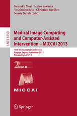
Medical Image Computing and Computer-Assisted Intervention — MICCAI 2013: 16th International Conference, Nagoya, Japan, September 22-26, 2013, Proceedings, Part II
دانلود کتاب پزشکی محاسبات تصویر پزشکی و مداخله به کمک کامپیوتر – MICCAI 2013: شانزدهمین کنگره بین المللی، ناگویا، ژاپن، 22-26 سپتامبر 2013، مجموعه مقالات، قسمت دوم
دسته: ابزار و تجهیزات, بیوشیمی, پزشکی, پیراپزشکی, عمومی, فناوری های تصویربرداری
| نویسنده |
Christian Barillot, Ichiro Sakuma, Kensaku Mori, Nassir Navab, Yoshinobu Sato |
|---|
| تعداد صفحهها |
718 |
|---|---|
| نوع فایل |
|
| حجم |
49 Mb |
| سال انتشار |
2013 |
89,000 تومان
دانلود ۳۰.۰۰۰ کتاب پزشکی فقط با قیمت یک کتاب و ۹۹ هزار تومان !
توضیحات
مجموعه سه جلدی LNCS 8149، 8150، و 8151 مراحل دادگاه شانزدهمین کنفرانس بین المللی محاسبات تصویر پزشکی و مداخله به کمک رایانه، MICCAI 2013، در ناگویا، ژاپن، در سپتامبر 2013 را تشکیل می دهد. بر اساس بررسی های دقیق همتایان، کمیته برنامه با دقت 262 مقاله اصلاح شده از 789 ارائه را برای ارائه در سه جلد انتخاب کرد. 86 مقاله موجود در جلد دوم در بخشهای موضوعی زیر سازماندهی شدهاند: ثبت و ساخت اطلس. میکروسکوپ، بافت شناسی و تشخیص به کمک کامپیوتر؛ مدل سازی حرکت و جبران. هش یادگیری ماشین، مدلسازی آماری و اطلسها؛ نشانگرهای زیستی؛ تشخیص و تصویربرداری به کمک رایانه؛ مدل سازی فیزیولوژیکی، شبیه سازی و برنامه ریزی؛ میکروسکوپ، تصویربرداری نوری و بافت شناسی. بیماری قلبی. رگ های خونی و ساختارهای لوله ای. تقسیم بندی مغز و اطلس. و کاربردهای کاربردی تشدید مغناطیسی و علوم اعصاب.
توضیحات(انگلیسی)
The three-volume set LNCS 8149, 8150, and 8151 constitutes the refereed proceedings of the 16th International Conference on Medical Image Computing and Computer-Assisted Intervention, MICCAI 2013, held in Nagoya, Japan, in September 2013. Based on rigorous peer reviews, the program committee carefully selected 262 revised papers from 789 submissions for presentation in three volumes. The 86 papers included in the second volume have been organized in the following topical sections: registration and atlas construction; microscopy, histology, and computer-aided diagnosis; motion modeling and compensation; segmentation; machine learning, statistical modeling, and atlases; computer-aided diagnosis and imaging biomarkers; physiological modeling, simulation, and planning; microscope, optical imaging, and histology; cardiology; vasculatures and tubular structures; brain segmentation and atlases; and functional MRI and neuroscience applications.


