مشاهده سبد خرید “Understanding Depression: Volume 1. Biomedical and Neurobiological Background 2018” به سبد خرید شما اضافه شد.
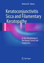
Keratoconjunctivitis Sicca and Filamentary Keratopathy: In Vivo Morphology in the Human Cornea and Conjunctiva 2013
دانلود کتاب پزشکی کراتوکونژونکتیویت سیکا و کراتوپاتی رشته ای: مورفوژنز in vivo در قرنیه و ملتحمه انسان.
دسته: پزشکی, پزشکی بالینی, چشم پزشکی
| نویسنده |
Helena M. Tabery |
|---|
| تعداد صفحهها |
196 |
|---|---|
| نوع فایل |
|
| حجم |
22 Mb |
| سال انتشار |
2013 |
89,000 تومان
دانلود ۳۰.۰۰۰ کتاب پزشکی فقط با قیمت یک کتاب و ۹۹ هزار تومان !
توضیحات
این کتاب تصاویری با بزرگنمایی بالا از دو بیماری ارائه میکند: کراتوکونژونکتیویت سیکا (KCS یا خشکی چشم)، یک بیماری بسیار شایع سطح چشم، و کراتوپاتی رشتهای، یک پدیده نسبتا نادر که معمولاً با KCS مرتبط است. تصاویر KCS طیف وسیعی از تغییرات سطح چشمی را نشان میدهند که در این بیماری مشاهده میشود، در حالی که تصاویر کراتوپاتی رشتهای به وضوح اجزای زائدههای سطح چشم، به اصطلاح بخیهها را نشان میدهند. عکسها پدیدههای ثبتشده در تنظیمات نوری مختلف، بدون رنگآمیزی و پس از رنگآمیزی با رنگهای تشخیصی را نشان میدهند و توالیهای عکاسی پویایی آنها را نشان میدهند. تصاویر منعکس کننده وضعیت in vivo هستند. هنگامی که از پدیده های مختلف آگاه شود، هر کسی که با تجهیزات تشخیصی استاندارد – لامپ شکاف و رنگ های تشخیصی کار می کند – می تواند تقریباً همه آنها را تشخیص دهد. این کتاب برای همه کسانی که با بیماری های سطح چشم سروکار دارند ارزشمند خواهد بود.
توضیحات(انگلیسی)
This book presents in vivo captured high-magnification images of two conditions: keratoconjunctivitis sicca (KCS, or dry eye), an extremely common disease of the ocular surface, and filamentary keratopathy, a relatively rare phenomenon most commonly associated with KCS. The images of KCS represent the broad spectrum of ocular surface changes seen in the condition while the images of filamentary keratopathy clearly reveal the components of the ocular surface appendices, termed filaments. The photographs show phenomena captured in various illumination modes, without staining and after staining with diagnostic dyes, and the photographic sequences illustrate their dynamics. The images reflect the in vivo situation. Once aware of the various phenomena, anyone working with standard diagnostic equipment - the slit lamp and the diagnostic dyes- will be able to detect almost all of them. The book will be invaluable for all who deal with ocular surface diseases.


