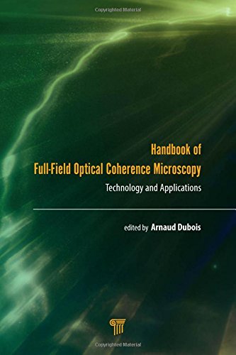
Handbook of Optical Coherence Microscopy: Technology and Applications 2015
دانلود کتاب پزشکی کتاب راهنمای میکروسکوپ انسجام نوری: فناوری و کاربردها
| نویسنده |
Arnaud Dubois |
|---|
| تعداد صفحهها |
800 |
|---|---|
| نوع فایل |
|
| حجم |
81 Mb |
| سال انتشار |
2015 |
89,000 تومان
میکروسکوپ انسجام نوری میدان کامل (FF-OCM) یک تکنیک تصویربرداری است که نماهای مقطعی از ریزساختار زیرسطحی اجسام نیمه شفاف را ارائه می دهد. این تکنیک مبتنی بر میکروسکوپ تداخلی با انسجام پایین است که از یک دوربین منطقهای برای تصویربرداری از چهره جسم روشنشده در میدان کامل استفاده میکند. FF-OCM از وضوح تصویربرداری جانبی میکروسکوپ نوری همراه با توانایی تقسیمبندی محوری نوری در دقت مقیاس میکرومتر سود میبرد. این فناوری را می توان در کاربردهای مختلف، به ویژه برای بررسی غیر تهاجمی بافت های بیولوژیکی استفاده کرد.
این راهنما اولین کتابی است که به طور کامل به FF-OCM اختصاص داده شده است. این در چهار بخش با مجموع 21 فصل که توسط کارشناسان شناخته شده و مشارکت کنندگان اصلی در این زمینه نوشته شده است، سازماندهی شده است. پس از مقدمهای کلی بر FF-OCM، ویژگیهای اساسی این فناوری مورد تجزیه و تحلیل و بحث تئوری قرار میگیرد. پیشرفت های کلیدی تکنولوژیکی FF-OCM برای بهبود سرعت گرفتن تصویر و تصویربرداری آندوسکوپی در قسمت دوم ارائه شده است. پسوندهای FF-OCM برای بهبود کنتراست تصویر یا تصویربرداری عملکردی در بخش سوم گزارش شده است. بخش آخر کتاب مروری بر کاربردهای بالقوه FF-OCM در پزشکی، زیست شناسی و علم مواد ارائه می دهد.
Full-field optical coherence microscopy (FF-OCM) is an imaging technique that provides cross-sectional views of the subsurface microstructure of semitransparent objects. The technology is based on low-coherence interference microscopy, which uses an area camera for en face imaging of the full-field illuminated object. FF-OCM benefits from the lateral imaging resolution of optical microscopy along with the capacity of optical axial sectioning at micrometer-scale resolution. The technique can be employed in diverse applications, in particular for non-invasive examination of biological tissues.
This handbook is the first to be entirely devoted to FF-OCM. It is organized into four parts with a total of 21 chapters written by recognized experts and major contributors to the field. After a general introduction to FF-OCM, the fundamental characteristics of the technology are analyzed and discussed theoretically. The main technological developments of FF-OCM for improving the image acquisition speed and for endoscopic imaging are presented in part II. Extensions of FF-OCM for image contrast enhancement or functional imaging are reported in part III. The last part of the book provides an overview of possible applications of FF-OCM in medicine, biology, and materials science.


