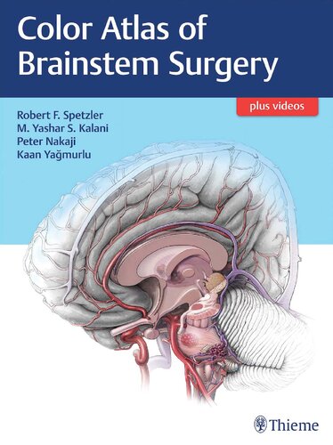
اطلس رنگی جراحی ساقه مغز ۲۰۱۷
Color Atlas of Brainstem Surgery 2017
دانلود کتاب اطلس رنگی جراحی ساقه مغز ۲۰۱۷ (Color Atlas of Brainstem Surgery 2017) با لینک مستقیم و فرمت pdf (پی دی اف)
| نویسنده |
Peter Nakaji, Robert F. Spetzler |
|---|
| تعداد صفحهها |
564 |
|---|---|
| نوع فایل |
epub, pdf |
| حجم |
115 Mb, 225 Mb |
| سال انتشار |
2017 |
89,000 تومان
معرفی کتاب اطلس رنگی جراحی ساقه مغز ۲۰۱۷
برنده جایزه اول جوایز کتاب پزشکی BMA 2018!
تخصص بسیار پیچیده جراحی ساقه مغز نیازمند سالها مطالعه، تاکید بر دقت و تعهد پرشور به تعالی است تا یک جراح مغز و اعصاب را برای مقابله با چالش های مهم تشریحی آماده کند. اگرچه ساقه مغز از نظر جراحی قابل دسترسی است، ساختار بسیار شیوای آن نیاز به تصمیمات جراحی دقیق دارد. درک عمیق ساقه مغز، آناتومی تالاموس و نواحی ورودی ایمن که برای دسترسی به نواحی حساس ساقه مغز استفاده می شود، برای پیمایش ایمن و موفق ساقه مغز ضروری است.
این اطلس جذاب و منحصر به فرد از تجربه چندین دهه نویسنده ارشد در انجام بیش از 1000 عمل جراحی بر روی ساقه مغز، تالاموس، عقده های قاعده ای و نواحی پری آپیکال استفاده می کند. محتوای آن بر اساس منطقه تشریحی سازماندهی شده است، و خوانندگان را قادر می سازد تا زیربخش های جداگانه ساقه مغز را مطالعه کنند، که هر کدام ملاحظات تشریحی و جراحی منحصر به فرد خود را دارند. این اطلس از روی جلد تا جلد، راهنمایی های فنی در مورد انتخاب رویکرد، زمان بندی جراحی و بهبود نتایج را که از طریق بیش از 1700 تصویر رنگی نفیس، آناتومی، عکس های بالینی، و نقاشی های خطی نشان داده شده است، در اختیار خوانندگان قرار می دهد.
نکات کلیدی
- برش ها و آناتومی مغز با جزئیات زیبا و بسیار توسعه یافته توسط کان یاگمورلو، که زیر نظر متخصص مغز و اعصاب و جراح مغز و اعصاب مشهور جهان آلبرت روتون جونیور آموزش دیده است.
- تصاویر رنگی که به وضوح با علامت گذاری ها و سایر نشانه های کانون های موردعلاقه برچسب گذاری شده اند، چندین نواحی ورود امن به ساقه مغز را شناسایی می کنند
- بیش از 50 مورد بیمار دقیق که تاریخچه هر بیمار از اختلالات عصبی قبلی، ارائه علائم و تصویربرداری قبل از عمل را برجسته می کند. روش های تشخیصی، جراحی برنامه ریزی شده، تعیین موقعیت بیمار، تصویربرداری حین و بعد از عمل و نتایج
- هفت انیمیشن و بیش از 50 فیلم جراحی که انتخاب رویکرد، یافته های آناتومیک و جراحی را برای ضایعات تالاموس و ساقه مغز نشان می دهد
ul>
این اطلس روشن بینشی را در مورد پیچیدگی های سالن های خاجی ساقه مغز فراهم می کند. جراحان مغز و اعصاب و جراحان مغز و اعصاب مقیم به طور یکسان که دانش را از مرواریدهای بالینی هر بخش استخراج می کنند، بدون شک جراحان ماهرتری خواهند شد تا به نفع بیماران خود باشند.
First Prize Winner at the 2018 BMA Medical Book Awards!
The highly complex specialty of brainstem surgery requires many years of study, a focus on precision, and a passionate dedication to excellence to prepare the neurosurgeon for navigating significant anatomic challenges. Although the brainstem is technically surgically accessible, its highly eloquent structure demands rigorous surgical decision-making. An in-depth understanding of brainstem and thalamic anatomy and the safe entry zones used to access critical areas of the brainstem is essential to traversing the brainstem safely and successfully.
This remarkable, one-of-a-kind atlas draws on the senior author's decades of experience performing more than 1,000 surgeries on the brainstem, thalamus, basal ganglia, and surrounding areas. Its content is organized by anatomic region, enabling readers to study separate subdivisions of the brainstem, each of which has its own unique anatomic and surgical considerations. From cover to cover, the atlas provides readers with technical guidance on approach selection, the timing of surgery, and optimization of outcomes-elucidated by more than 1700 remarkable color illustrations, dissections, clinical images, and line drawings.
Key Highlights
- Beautifully detailed, highly sophisticated brain slices and dissections by Kaan Yagmurlu, who trained under the internationally renowned neuroanatomist and neurosurgeon Albert Rhoton Jr.
- Color illustrations clearly labeled with callouts and other indicators of foci of interest delineate multiple safe entry zones to the brainstem
- More than 50 detailed patient cases highlight each patient's history of previous neurological disorders, presenting symptoms, preoperative imaging, diagnosis, the planned surgical approach, patient positioning, intraoperative and postoperative imaging, and outcome
- Seven animations and more than 50 surgical videos elucidate approach selection, anatomy, and surgical outcomes of thalamic region and brainstem lesions
This illuminating atlas provides insights into the complexities of the hallowed halls of the brainstem. Neurosurgeons and neurosurgical residents alike who glean knowledge from the clinical pearls throughout each section will no doubt become more adept surgeons, to the ultimate benefit of their patients.


