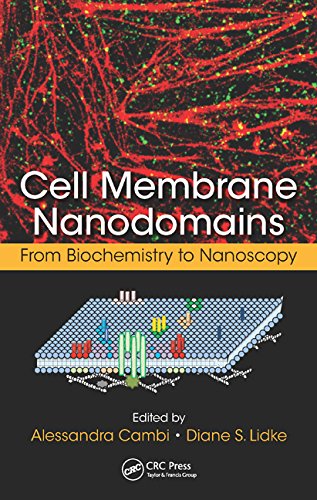
نانودامنههای غشای سلولی: از بیوشیمی تا نانوسکوپی، پیشرفتهای اخیر در درک ما از سازماندهی غشا را با تمرکز ویژه بر تکنیکهای تصویربرداری پیشرفته که این اکتشافات جدید را ممکن میسازد، توصیف میکند. با مشارکت پیشگامان در این زمینه، این کتاب به بررسی زمینه هایی می پردازد که در آن کاربرد این فناوری های جدید مفاهیم جدیدی را در زیست شناسی نشان می دهد. این مجموعه ای از کار را گرد هم می آورد که در آن ادغام زیست شناسی غشایی و میکروسکوپ بر ماهیت بین رشته ای این زمینه هیجان انگیز تأکید می کند.
این کتاب با توصیف گسترده ای از سازماندهی غشاء، از جمله کار ضروری بر روی پارتیشن بندی لیپیدها در سیستم های مدل و نقش پروتئین ها در سازماندهی غشا، به بررسی چگونگی تقسیم لیپید و تنظیم غشا می تواند عملکرد پروتئین و انتقال سیگنال را تنظیم کند. . سپس بر پیشرفتهای اخیر در تکنیکها و ابزارهای تصویربرداری تمرکز میکند که پیشرفتهای بیشتری را در درک ما از نانوپلتفرمهای سیگنالینگ پیش میبرد. پوشش شامل چندین تکنیک تصویربرداری محدود با پراش است که امکان اندازهگیری توزیع/کلوپ شدن پروتئین و انحنای غشاء در سلولهای زنده، پروتئینهای فلورسنت جدید، آنالیزهای جدید Lourdan و جعبه ابزاری از احتمالات برچسبگذاری با استفاده از رنگهای آلی را فراهم میکند.
از آنجایی که تکنیکهای نوری با وضوح فوقالعاده برای پیشرفت درک ما از ساختار سلولی و رفتار پروتئین ضروری بوده است، این کتاب با بحث در مورد فنآوریهایی که امکان تجسم لیپیدها، پروتئینها و سایر اجزای مولکولی را فراهم میکند، به پایان میرسد. تفکیک مکانی و زمانی بی سابقه همچنین نکات و نکات حوزه به سرعت در حال تکامل میکروسکوپ با وضوح بالا، از جمله روشهای جدید و ابزارهای تجزیه و تحلیل دادهها را که منحصراً به این تکنیکها مربوط میشوند، توضیح میدهد.
ادغام زیست شناسی غشایی و فناوری های تصویربرداری پیشرفته بر ماهیت چند رشته ای این زمینه هیجان انگیز تأکید می کند. طیف وسیعی از مشارکتهای کارشناسان برجسته بینالمللی، این کتاب را به ابزاری ارزشمند برای تجسم سیگنالهای نانوپلتفرمها با استفاده از ابزارهای میکروسکوپ نوری پیشرفته تبدیل کرده است.
Cell Membrane Nanodomains: From Biochemistry to Nanoscopy describes recent advances in our understanding of membrane organization, with a particular focus on the cutting-edge imaging techniques that are making these new discoveries possible. With contributions from pioneers in the field, the book explores areas where the application of these novel techniques reveals new concepts in biology. It assembles a collection of works where the integration of membrane biology and microscopy emphasizes the interdisciplinary nature of this exciting field.
Beginning with a broad description of membrane organization, including seminal work on lipid partitioning in model systems and the roles of proteins in membrane organization, the book examines how lipids and membrane compartmentalization can regulate protein function and signal transduction. It then focuses on recent advances in imaging techniques and tools that foster further advances in our understanding of signaling nanoplatforms. The coverage includes several diffraction-limited imaging techniques that allow for measurements of protein distribution/clustering and membrane curvature in living cells, new fluorescent proteins, novel Laurdan analyses, and the toolbox of labeling possibilities with organic dyes.
Since superresolution optical techniques have been crucial to advancing our understanding of cellular structure and protein behavior, the book concludes with a discussion of technologies that are enabling the visualization of lipids, proteins, and other molecular components at unprecedented spatiotemporal resolution. It also explains the ins and outs of the rapidly developing high- or superresolution microscopy field, including new methods and data analysis tools that exclusively pertain to these techniques.
This integration of membrane biology and advanced imaging techniques emphasizes the interdisciplinary nature of this exciting field. The array of contributions from leading world experts makes this book a valuable tool for the visualization of signaling nanoplatforms by means of cutting-edge optical microscopy tools.


