راهنمای مصور بافت شناسی دهان ۲۰۲۱
An Illustrated Guide to Oral Histology 2021
دانلود کتاب راهنمای مصور بافت شناسی دهان ۲۰۲۱ (An Illustrated Guide to Oral Histology 2021) با لینک مستقیم و فرمت pdf (پی دی اف)
| نویسنده |
Imran Farooq, Paul Anderson, Saqib Ali |
|---|
ناشر:
Wiley
دسته: پزشکی, پزشکی عمومی, جراحی دهان, دندانپزشکی

۳۰ هزار تومان تخفیف با کد «OFF30» برای اولین خرید
| سال انتشار |
2021 |
|---|---|
| زبان |
English |
| تعداد صفحهها |
192 |
| نوع فایل |
|
| حجم |
6 Mb |
🏷️ 200,000 تومان قیمت اصلی: 200,000 تومان بود.129,000 تومانقیمت فعلی: 129,000 تومان.
🏷️
378,000 تومان
قیمت اصلی: ۳۷۸٬۰۰۰ تومان بود.
298,000 تومان
قیمت فعلی: ۲۹۸٬۰۰۰ تومان.
📥 دانلود نسخهی اصلی کتاب به زبان انگلیسی(PDF)
🧠 به همراه ترجمهی فارسی با هوش مصنوعی
🔗 مشاهده جزئیات
دانلود مستقیم PDF
ارسال فایل به ایمیل
پشتیبانی ۲۴ ساعته
توضیحات
معرفی کتاب راهنمای مصور بافت شناسی دهان ۲۰۲۱
راهنمای مصور بافت شناسی دهان
مقدمه ای بر تکامل دندان، شامل مراحل جوانه، کلاهک، زنگ اولیه و زنگ متاخر
بررسی کامل مینا، عاج، سمنتوم و پالپ دندان
بحث در مورد رباط پریودنتال، شامل الیاف تاج آلوئولی، الیاف افقی، مایل، اپیکال و بین ریشه ای، الیاف ترانس سپتال و الیاف لثه ای
راهنمای استخوان آلوئولار، مخاط دهان و غدد بزاقی
با استفاده از این مرجع دندانپزشکی مصور با کیفیت بالا، اطلاعات بیشتری در مورد ارائه بافت شناسی بافت های دهانی بیمار و سالم کسب کنید.
راهنمای مصور بافت شناسی دهان مجموعه ای از تصاویر بافت شناسی و آسیب شناسی با کیفیت بالا ارائه می کند که بافت های دهانی بیمار و سالم را به تصویر می کشد.
این کتاب بیش از 200 میکروگراف با بزرگنمایی بالا از بافت های دهان، و همچنین تعاریف و توضیحاتی در مورد ویژگی های کلیدی شناسایی بافت شناسی و آسیب شناسی بافت های دهان ارائه می دهد. خوانندگان همچنین از توضیحات اهمیت بالینی ویژگی های خاص، تصاویر متعددی از مقاطع سنگ زده، مقاطع رنگ آمیزی شده با هماتوکسیلین و ائوزین، و تصاویر الکترونی بهره مند خواهند شد. این کتاب همچنین شامل مباحث اصلی مانند موارد زیر است:
راهنمای مصور بافت شناسی دهان که برای دانشجویان تحصیلات تکمیلی دندانپزشکی عالی است، برای دانشجویان مقطع کارشناسی دندانپزشکی و کسانی که به دنبال بهبود درک خود از ساختار میکروسکوپی بافت های دندانی و آسیب شناسی های آنها هستند نیز مفید خواهد بود.
توضیحات(انگلیسی)
An Illustrated Guide to Oral Histology
An introduction to tooth development, including the bud, cap, early bell, and late bell stages
A thorough exploration of enamel, dentin, cementum and dental pulp
A discussion of the periodontal ligament, including alveolar crest fibers, horizontal, oblique, apical, and inter-radicular fibers, transseptal fibers, and gingival fibers
A guide to alveolar bone, oral mucosa, and salivary glands
Learn more about the histological presentation of diseased and normal oral tissues with this high definition illustrated dental reference
An Illustrated Guide to Oral Histology delivers a collection of high-definition histological and pathological images, presenting both diseased and normal oral tissues.
The book provides over 200 high-magnification histomicrographs of oral tissues, as well as definitions and explanations of key identifying histological and pathological features of oral tissues. Readers will also benefit from explanations of the clinical significance of particular features, numerous images of ground sections, haemotoxylin- and eosin-stained sections, and electron images. It also includes core topics such as:
Perfect for postgraduate dental students, An Illustrated Guide to Oral Histology will also be useful to undergraduate dental students, and those looking to improve their understanding of the microscopic structure of dental tissues and their pathologies.
دیگران دریافت کردهاند
راهنمای مصور پرواز ۲۰۲۲
An Illustrated Guide to Flying 2022
🏷️ 200,000 تومان قیمت اصلی: 200,000 تومان بود.129,000 تومانقیمت فعلی: 129,000 تومان.
راهنمای مصور بیماری های مادرزادی قلب: از تشخیص تا درمان – از جنین تا بزرگسالی ۲۰۱۹
An Illustrated Guide to Congenital Heart Disease: From Diagnosis to Treatment – From Fetus to Adult 2019
🏷️ 200,000 تومان قیمت اصلی: 200,000 تومان بود.129,000 تومانقیمت فعلی: 129,000 تومان.
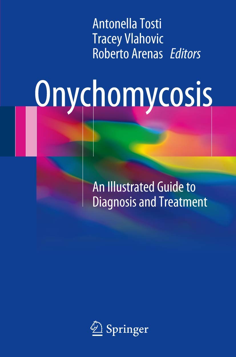
اونیکومایکوزیس: راهنمای مصور تشخیص و درمان ۲۰۱۷
Onychomycosis: An Illustrated Guide to Diagnosis and Treatment 2017
🏷️ 200,000 تومان قیمت اصلی: 200,000 تومان بود.129,000 تومانقیمت فعلی: 129,000 تومان.
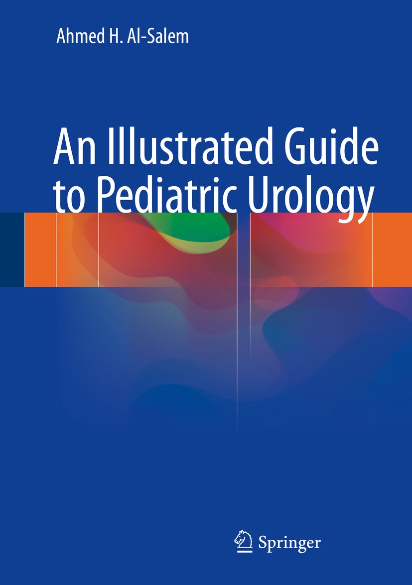
راهنمای مصور اورولوژی اطفال ۲۰۱۶
An Illustrated Guide to Pediatric Urology 2016
🏷️ 200,000 تومان قیمت اصلی: 200,000 تومان بود.129,000 تومانقیمت فعلی: 129,000 تومان.
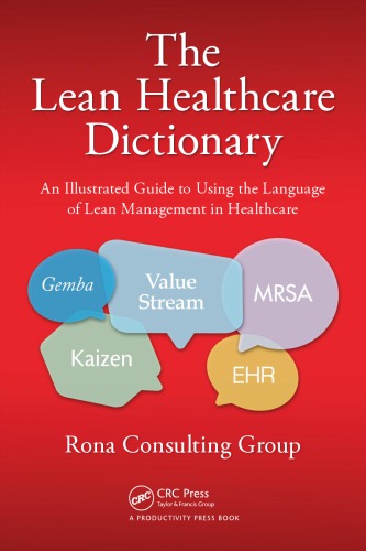
فرهنگ لغت لین در حوزه سلامت: راهنمای مصور استفاده از زبان مدیریت لین در مراقبت های بهداشتی ۲۰۱۴
The Lean Healthcare Dictionary: An Illustrated Guide to Using the Language of Lean Management in Healthcare 2014
🏷️ 200,000 تومان قیمت اصلی: 200,000 تومان بود.129,000 تومانقیمت فعلی: 129,000 تومان.
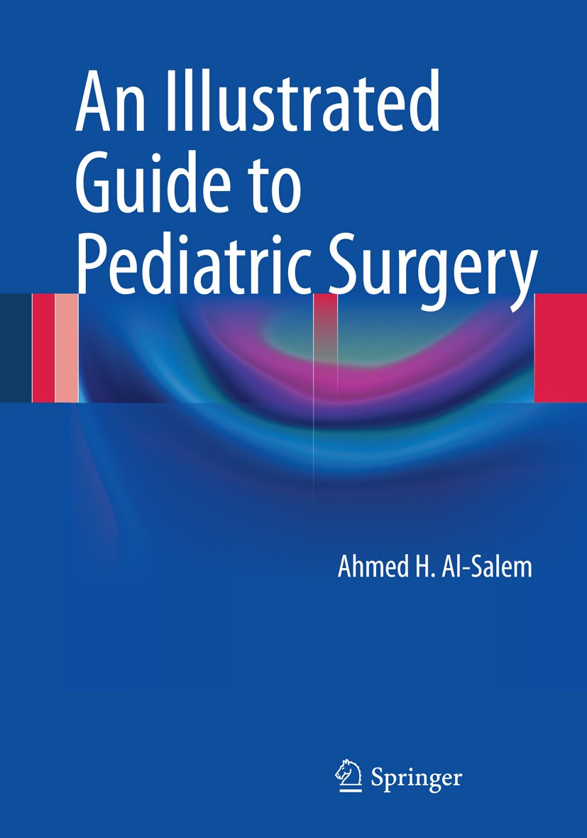
راهنمای مصور جراحی کودکان ۲۰۱۴
An Illustrated Guide to Pediatric Surgery 2014
🏷️ 200,000 تومان قیمت اصلی: 200,000 تومان بود.129,000 تومانقیمت فعلی: 129,000 تومان.
سایر کتابهای ناشر
ویلی ۲۰۲۲ تفسیر و کاربرد استانداردهای IFRS ۲۰۲۲
Wiley 2022 Interpretation and Application of IFRS Standards 2022
🏷️ 200,000 تومان قیمت اصلی: 200,000 تومان بود.129,000 تومانقیمت فعلی: 129,000 تومان.
Wiley GAAP: دفترچه راهنمای افشای صورتهای مالی ۲۰۲۱
Wiley GAAP: Financial Statement Disclosure Manual 2021
🏷️ 200,000 تومان قیمت اصلی: 200,000 تومان بود.129,000 تومانقیمت فعلی: 129,000 تومان.
کتابچه راهنمای Wiley از آنچه در مدیریت ریسک خشونت کار می کند: تئوری ، تحقیق و تمرین ۲۰۱۹
The Wiley Handbook of What Works in Violence Risk Management: Theory, Research, and Practice 2019
🏷️ 200,000 تومان قیمت اصلی: 200,000 تومان بود.129,000 تومانقیمت فعلی: 129,000 تومان.
Wiley IFRS 2017: تفسیر و کاربرد استانداردهای IFRS ۲۰۱۷
Wiley IFRS 2017: Interpretation and Application of IFRS Standards 2017
🏷️ 200,000 تومان قیمت اصلی: 200,000 تومان بود.129,000 تومانقیمت فعلی: 129,000 تومان.
Wiley IFRS 2016: تفسیر و کاربرد استانداردهای بینالمللی گزارشگری مالی ۲۰۱۶
Wiley IFRS 2016: Interpretation and Application of International Financial Reporting Standards 2016
🏷️ 200,000 تومان قیمت اصلی: 200,000 تومان بود.129,000 تومانقیمت فعلی: 129,000 تومان.
Wiley GAAP: راهنمای کاربردی و دفتر تمرین ۲۰۱۰
Wiley GAAP: Practical Implementation Guide and Workbook 2010
🏷️ 200,000 تومان قیمت اصلی: 200,000 تومان بود.129,000 تومانقیمت فعلی: 129,000 تومان.
✨ ضمانت تجربه خوب مطالعه
بازگشت کامل وجه
در صورت مشکل، مبلغ پرداختی بازگردانده می شود.
دانلود پرسرعت
دانلود فایل کتاب با سرعت بالا
ارسال فایل به ایمیل
دانلود مستقیم به همراه ارسال فایل به ایمیل.
پشتیبانی ۲۴ ساعته
با چت آنلاین و پیامرسان ها پاسخگو هستیم.
ضمانت کیفیت کتاب
کتاب ها را از منابع معتیر انتخاب می کنیم.

