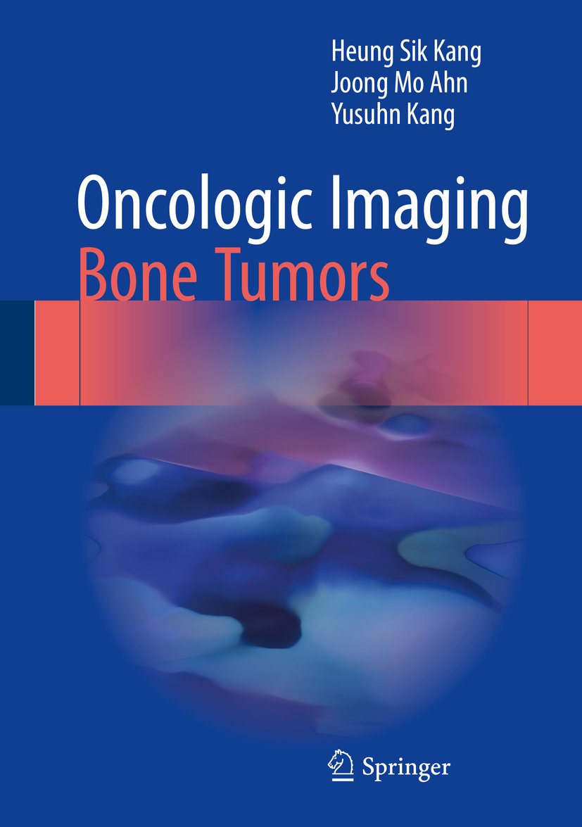
تصویربرداری انکولوژیک: تومورهای استخوان ۲۰۱۷
Oncologic Imaging: Bone Tumors 2017
دانلود کتاب تصویربرداری انکولوژیک: تومورهای استخوان ۲۰۱۷ (Oncologic Imaging: Bone Tumors 2017) با لینک مستقیم و فرمت pdf (پی دی اف)
| نویسنده |
Heung Sik Kang, Joong Mo Ahn, Yusuhn Kang |
|---|
ناشر:
Springer

۳۰ هزار تومان تخفیف با کد «OFF30» برای اولین خرید
| سال انتشار |
2017 |
|---|---|
| زبان |
English |
| تعداد صفحهها |
382 |
| نوع فایل |
|
| حجم |
48 Mb |
🏷️ 200,000 تومان قیمت اصلی: 200,000 تومان بود.129,000 تومانقیمت فعلی: 129,000 تومان.
🏷️
378,000 تومان
قیمت اصلی: ۳۷۸٬۰۰۰ تومان بود.
298,000 تومان
قیمت فعلی: ۲۹۸٬۰۰۰ تومان.
📥 دانلود نسخهی اصلی کتاب به زبان انگلیسی(PDF)
🧠 به همراه ترجمهی فارسی با هوش مصنوعی
🔗 مشاهده جزئیات
دانلود مستقیم PDF
ارسال فایل به ایمیل
پشتیبانی ۲۴ ساعته
توضیحات
معرفی کتاب تصویربرداری انکولوژیک: تومورهای استخوان ۲۰۱۷
هدف این کتاب، ارائه درکی صحیح از طیف تظاهرات رادیولوژیک تومورهای استخوانی به خوانندگان است، تظاهراتی که بازتاب دهنده هیستوپاتولوژی و آنالیز الگوهای یافته های تصویربرداری هستند. بخش اول کتاب، مفاهیم اساسی و پارامترهای تشخیصی را توضیح می دهد، از جمله جمعیت شناسی، محل ضایعه، فعالیت بیولوژیکی، معدنی شدن ماتریکس و واکنش های اندوستئال و پریوستیال. در بخش دوم، ویژگی های رادیولوژیک تیپیک و آتیپیک تومورهای استخوانی با جزئیات بررسی می شوند، با تاکید بر یافته های رادیوگرافی و ام آرآی مشخصه و ارجاع به طرح های شماتیک و تصاویر پاتولوژیک یا حین عمل در صورت لزوم. بخش سوم بر حل مسئله در مواردی که در عمل رادیولوژی واقعی با آن ها مواجه می شویم متمرکز است و الگوهای قطعی را بر اساس متغیرهای رادیوگرافی و ام آرآی و محل ضایعه مشخص می کند. موارد آموزنده تصویرسازی و مقایسه شده اند تا درک تشخیص افتراقی با استفاده از آنالیز الگو را بهبود بخشند. بخش پایانی کتاب به خوانندگان کمک می کند تا آنچه را که آموخته اند تثبیت کنند و با ارائه حدود 30 مورد تیپیک تومور استخوانی همراه با سوالات، پاسخ ها و توضیحات، مهارت های استدلال تشخیصی خود را تقویت کنند.
توضیحات(انگلیسی)
This book aims to provide readers with a sound understanding of the spectrum of radiologic appearances of bone tumors, which reflect histopathology, and the pattern analysis of imaging findings.The first part of the book explains basic concepts and diagnostic parameters, including demographics, lesion location, biological activity, matrix mineralization, and endosteal and periosteal reactions. In the second part, typical and atypical radiologic features of bone tumors are reviewed in detail, with emphasis on the characteristic radiographic and MR imaging findings and reference to schematic drawings and pathologic or operative images when appropriate.The third part focuses on problem solving in cases encountered in real radiology practice and identifies categorical patterns on the basis of radiographic and MR variables and lesion location. Informative cases are illustrated and compared to enhance understanding of differential diagnosis using pattern analysis. The final part of the book helps readers to consolidate what they have learned and to hone their diagnostic reasoning skills by presenting about 30 typical bone tumor cases with questions, answers, and commentary.
دیگران دریافت کردهاند
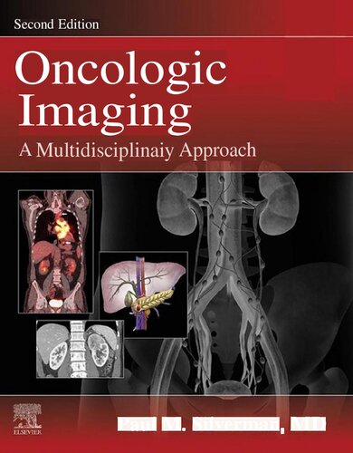
تصویربرداری انکولوژیک: یک رویکرد چند رشته ای ۲۰۲۲
Oncologic Imaging: A Multidisciplinary Approach 2022
طب داخلی, انکولوژی, پزشکی, پزشکی عمومی, تصویربرداری تشخیصی, جراحی, رادیولوژی، رادیوتراپی و پزشکی هسته ای
🏷️ 200,000 تومان قیمت اصلی: 200,000 تومان بود.129,000 تومانقیمت فعلی: 129,000 تومان.
تصویربرداری انکولوژیک: تومورهای استخوان ۲۰۱۷
Oncologic Imaging: Bone Tumors 2017
انکولوژی, بیوشیمی پزشکی, پاتولوژی, پزشکی, پزشکی بالینی, پزشکی عمومی, پیراپزشکی, فناوری های تصویربرداری
🏷️ 200,000 تومان قیمت اصلی: 200,000 تومان بود.129,000 تومانقیمت فعلی: 129,000 تومان.
تصویربرداری انکولوژیک: تومورهای ستون فقرات و نخاع، ۲۰۱۷
Oncologic Imaging: Spine and Spinal Cord Tumors 2017
انکولوژی, بیوشیمی پزشکی, پاتولوژی, پزشکی, پزشکی بالینی, پزشکی عمومی, پیراپزشکی, فناوری های تصویربرداری
🏷️ 200,000 تومان قیمت اصلی: 200,000 تومان بود.129,000 تومانقیمت فعلی: 129,000 تومان.
تصویربرداری انکولوژیک: اورولوژی ۲۰۱۶
Oncologic Imaging: Urology 2016
انکولوژی, بیوشیمی پزشکی, پاتولوژی, پزشکی, پزشکی بالینی, پزشکی عمومی, پیراپزشکی, جراحی, فناوری های تصویربرداری
🏷️ 200,000 تومان قیمت اصلی: 200,000 تومان بود.129,000 تومانقیمت فعلی: 129,000 تومان.
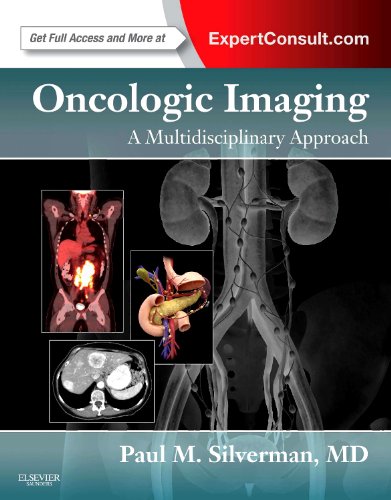
تصویربرداری سرطان شناسی: یک رویکرد چندرشته ای ۲۰۱۲
Oncologic Imaging: A Multidisciplinary Approach 2012
🏷️ 200,000 تومان قیمت اصلی: 200,000 تومان بود.129,000 تومانقیمت فعلی: 129,000 تومان.
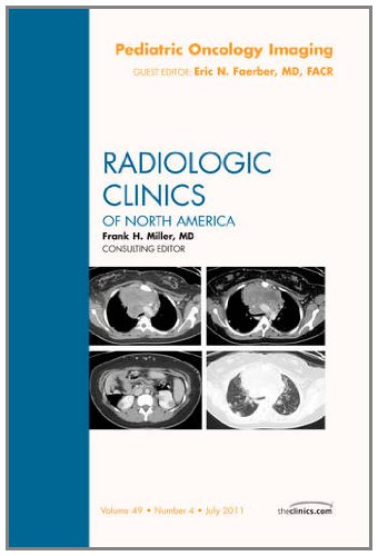
تصویربرداری سرطان شناسی کودکان ۲۰۱۱
Pediatric Oncology Imaging 2011
🏷️ 200,000 تومان قیمت اصلی: 200,000 تومان بود.129,000 تومانقیمت فعلی: 129,000 تومان.
سایر کتابهای ناشر
راهنمای اسپرینگر در زمینه بیوتکنولوژی دریایی ۲۰۱۵
Springer Handbook of Marine Biotechnology 2015
🏷️ 200,000 تومان قیمت اصلی: 200,000 تومان بود.129,000 تومانقیمت فعلی: 129,000 تومان.
اسپرینتر و اسپرامر ۲۰۱۴
Sprinter and Sprummer 2014
🏷️ 200,000 تومان قیمت اصلی: 200,000 تومان بود.129,000 تومانقیمت فعلی: 129,000 تومان.
راهنمای اسپرینگر در زمینهٔ آگاهی زیستی/آگاهی عصبی ۲۰۱۳
Springer Handbook of Bio-/Neuro-Informatics 2013
🏷️ 200,000 تومان قیمت اصلی: 200,000 تومان بود.129,000 تومانقیمت فعلی: 129,000 تومان.
✨ ضمانت تجربه خوب مطالعه
بازگشت کامل وجه
در صورت مشکل، مبلغ پرداختی بازگردانده می شود.
دانلود پرسرعت
دانلود فایل کتاب با سرعت بالا
ارسال فایل به ایمیل
دانلود مستقیم به همراه ارسال فایل به ایمیل.
پشتیبانی ۲۴ ساعته
با چت آنلاین و پیامرسان ها پاسخگو هستیم.
ضمانت کیفیت کتاب
کتاب ها را از منابع معتیر انتخاب می کنیم.

